Over the last 4 decades many new methods and instruments for objective assessment of the skin have been brought to the market. In an attempt to categorize, one can identify these developments as single-point methods, 2D imaging methods (top view or cross-sectional) and, more recently, full 3D imaging methods. Some methods further allow for investigation of dynamic changes in skin parameters with the use of both time plot graphs and real time or compressed time scale video-clips. Most methods are non-invasive in nature and incorporate both contact and non-contact measurement technologies.
Tissue Viability Imaging for quantitative user-independent assessment
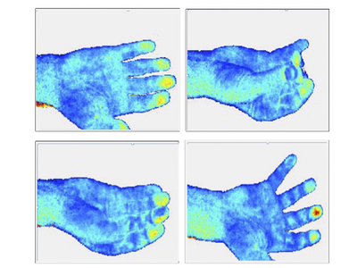
This report looks at the Tissue Viability Imaging for quantitative user-independent assessment of erythema, blanching and other skin parameters
You need to be a subscriber to read this article.
Click here to find out more.
Click here to find out more.
You may also like
Digital
![De-Dupe the Copycats: Protecting beauty innovation and consumer safety on TikTok Shop]()

De-Dupe the Copycats: Protecting beauty innovation and consumer safety on TikTok Shop
Read moreMeet the speakers of SnapDragon's dedicated Beauty and Personal Care Industry LinkedIn Live webinar on 1st July: De-Dupe the Copycats. The online webinar will bring together three perspectives that rarely sit in the same room: platform enforcement, brand protection strategy and cosmetic science
Trending Articles
You may also like
Digital
![De-Dupe the Copycats: Protecting beauty innovation and consumer safety on TikTok Shop]()
De-Dupe the Copycats: Protecting beauty innovation and consumer safety on TikTok Shop
Meet the speakers of SnapDragon's dedicated Beauty and Personal Care Industry LinkedIn Live webinar on 1st July: De-Dupe the Copycats. The online webinar will bring together three perspectives that rarely sit in the same room: platform enforcement, brand protection strategy and cosmetic science
Digital
![Haut.AI Collaborates with OLAY on Virtual Companion Technology to Power Clinically Modeled Skincare Simulations]()

Haut.AI Collaborates with OLAY on Virtual Companion Technology to Power Clinically Modeled Skincare Simulations
Haut.AI today announced its collaboration with OLAY, a Procter & Gamble (P&G) brand, to introduce an innovative Virtual Companion technology within the OLAY Skin Advisor experience

You need to be a subscriber to read this article.
Click here to find out more.
Click here to find out more.


Digital
![Haut.AI Brings AI Skin Intelligence to VivaTech 2026]()

Haut.AI Brings AI Skin Intelligence to VivaTech 2026
Haut.AI, an award-winning company specialising in AI-powered skin intelligence, will be exhibiting at VivaTech 2026 in Paris. Deployed across 130+ brand and retail partners worldwide, its platform will be on show at Booth 2H06-006 from June 17 to 20, with live demonstrations of AI Skin Analysis,nSkinGPT, and Skin.Chat
